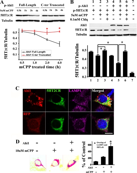
FIGURE 2.
Ahi1 promoted the degradation of 5-HT2CR through the lysosomal pathway. A, full-length or truncated Ahi1 was co-transfected with 5-HT2CR into N18TG2 cells, and then immunoblotting was performed. In the cells transfected with full-length Ahi1, 5-HT2CR decreased in a time-dependent way after mCPP treatment, whereas in the cells transfected with truncated Ahi1 this decrease was inhibited. B, co-expression of 5HT2CR with Ahi1 in N18TG2 cells led to increased degradation of mCPP-activated 5-HT2CR compared with the expression of 5-HT2CR alone (lane 5 compared with lane 2), which could be blocked under the application of Chlq (lane 6). C, 5-HT2CR located in the lysosomes was increased in the presence of Ahi1. In cells expressing GFP-5-HT2CR and RFP-Ahi1, the 5-HT2CR mainly overlapped with the lysosomes (LAMP1 as marker) and co-localized with Ahi1 (top row, indicated by arrows). In the absence of Ahi1, 5-HT2CR is distributed throughout the plasma membrane as well as the cytoplasm (bottom row). D, quantifying the co-localization of 5-HT2CR with the lysosomes in N18TG2 cells by calculating the area containing 5-HT2CR and lysosome-labeled pixels with or without Ahi1. Scale bar: 10 μm. Data are expressed as mean ± S.E. *, statistically different from control group. C-ter, C terminus; p-Ahi1, plasmid-Ahi1; p-5HT2CR, plasmid-5HT2CR; LAMP1, lysosome-associated membrane protein 1; RFP, red fluorescent protein.

