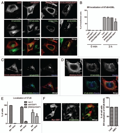
Figure 9.
Effect of recombinant ArfGAP1 on COPI dependent retrograde transport. COPI dependent transport was measured using STxB-KDEL conjugated with Alexa 488. (A) Relative distribution of STxB-KDEL and ArfGAP1-myc. STxB-KDEL conjugated with Alexa 488 fluorescence was internalized for 3hr in HeLa cells transfected with indicated constructs. After fixation, cells were stained with anti-myc antibody (red). Representative images are shown. (B) Quantitation of STxB-KDEL targeting to the ER. The percentage of transfected cells with a reticular distribution consistent with an ER localization of STxB-KDEL (referred to as ER localization) was determined. More than 50 cells were counted, and the summary of 3 independent experiments is shown. Data for the 3 h time point were analyzed by one way ANOVA followed by the Bonferoni multiple comparisons test. * indicates p < 0.05 compared with LacZ. (C) Distribution of STxB-KDEL in cells expressing [CC22,25SS]ArfGAP1. After internalization of STxB-KDEL (green) for 3 h, cells expressing [CC22,25SS]ArfGAP1 were stained for the Golgi marker giantin (blue) and [CC22,25SS]ArfGAP1 (red). STxB-KDEL localized at the perinuclear region, partially colocalizing with giantin but also was present in a structure connected to the Golgi. [CC22,25SS] ArfGAP1 colocalized with STxB-KDEL in the latter structure. (D) Distribution of STxB-KDEL in cells expressing [R50K]ArfGAP1. Cells expressing [R50K]ArfGAP1 were treated as described in C. The punctuate structure was also stained by giantin. (E) Quantification of relative reticular and punctate distributions illustrated in (D). The cells with STxB-KDEL localization that was reticular (presumed to be ER) or ER and puncta (dots) were shown as the percentage of transfected cells. Summary of 3 independent experiments is shown. Data were analyzed by two way ANOVA followed by the Bonferoni multiple comparisons test. * indicates p < 0.05 compared with Lac Z control. (F) Relative distribution of STxB-KDEL and GBF1-myc, and quantification of STxB-KDEL localization. STxB-KDEL was internalized as above, and ER localization of STxB-KDEL is shown as the percentage of transfected cells.

