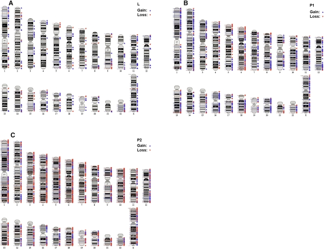
Figure 5.
Chromosomal abnormalities in plasma preceding relapse. CNVs based on 50 consecutive markers (SNP and/or CN) and a minimum segment size of 50,000 bp. Example of array karyotypes of cfDNA for one patient preceding relapse: (A) normal leukocyte DNA sample, (B) P1 cfDNA sample taken 6 yr after diagnosis, and (C) P2 cfDNA taken 1 mo before the patient was diagnosed with metastatic disease. There was a significant increase in CNVs detected between P1 and P2: P1, 387 (79.08%) amplifications and 96 (20.92%) deletions; P2, 1332 (53.67%) amplifications and 1150 (46.33%) deletions.

