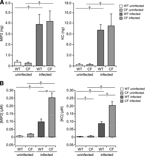
Figure 5.
Total chemokine secretion and concentrations of WT and ΔF508 CF mTECs, 24 hours after stimulation with P. aeruginosa PAO1. (a) Total MIP-2 and KC released from WT and ΔF508 CF mTECs, before and 24 hours after infection with 108 CFU/ml P. aeruginosa PAO1 (n = 5–7 Transwells). No differences were evident in patterns or quantities of chemokines released from WT and ΔF508 CF mTECs. (b) Apical and basolateral concentrations of MIP-2 and KC were derived, using calculated apical surface volumes for WT and ΔF508 CF mTECs before and 24 hours after infection with 108 CFU/ml P. aeruginosa PAO1 (n = 5–7 Transwells). Means ± SEM are shown. *P ≤ 0.05 and †P ≤ 0.005, using unpaired Student t tests for statistical comparisons. Although CXC-chemokines were primarily secreted basolaterally, greater concentrations were present at the apical surface because of the minute volume of surface-lining fluid. The apical microenvironment of airway epithelial cells amplifies the inflammatory signal, which is greater at the surface of CF epithelia.

