
Figure 5.
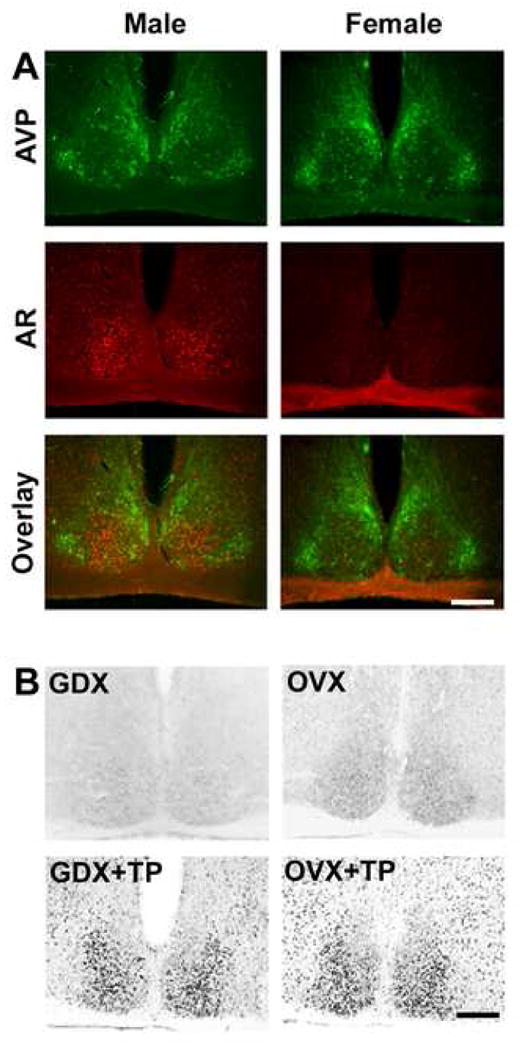
(A) Photomicrographs show fluorescent double-labeled AR- (red) and AVP-ir (green) in SCN of intact male and female mice. Note that AR-ir is localized to the core SCN region lacking AVP-ir. Upper panel shows AVP, middle panel shows AR, and lower panel shows overlay. (B) Photomicrographs show AR-ir in GDX/OVX (upper panel) and GDX/OVX+TP (lower panel) treated male and female mice.