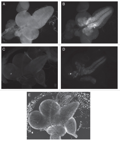
Figure 4.
Immunolocalization of ER and E(spl) in the 3rd instar larval CNS and optic lobes. (A and B) e(r)+ male, exposure 100 msec. (C and D) e(r)27-1 male, exposure 250 msec. (E) DAPI staining of (C and D). This aids in the identification of the tissues. (A and C) ER immunostaining. (B and D) E(spl) immunostaining. (A) ER immunostaining is seen throughout the larval CNS and optic lobes, (B) E(spl) is seen in a subset of cells in the larval CNS. (C) ER is vastly reduced in an e(r)27-1 mutant. (D) In the same e(r)27-1 mutant, E(spl) staining is also clearly reduced when compared to wild-type, even with 2.5 times the exposure.

