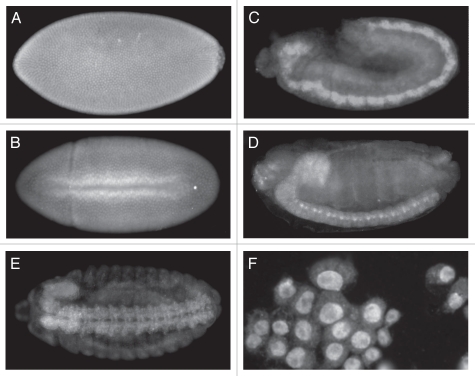
Figure 5.
Immunolocalization of ER in wild-type embryos and Schneider 2 cells. Anterior is to the left and posterior is to the right for the embryos. (A) Stage 5-Syncytial blastoderm showing pole cells on the right. ER is localized to the nuclei. (B) Stage 7-ventral mesoderm invagination. ER can still be seen localized primarily to the nuclei. (C) Stage 11. ER shows a diffuse general localization, but much higher levels in the developing CNS. (D) Stage 14. Staining is seen in the brain lobe on the anterior dorsal side of the embryo and in the ventral nerve cord. (E) Stage 14, ventral view. Staining is seen in the brain lobe and the two tracks of the ventral nerve cord. Staining also appears in the peripheral nervous system, although at lower levels. (F) Schneider 2 cells. Staining is clearly localized to the nuclei.

