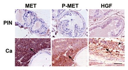
Figure 4.
Expression (arrows) of total (MET) and phosphorylated (P-MET) MET and HFGα (HGF) in prostate neoplastic lesions. Carcinomas (Ca) and prostate intraepithelial neoplasms (PIN), are located, respectively, in the proximal and distal regions of prostatic ducts of mice with prostate epithelium-specific inactivation of p53 and Rb. Note absence of MET expression in PIN and HGFα location in stromal (arrow) but not epithelial (arrowhead) cells. ABC Elite method, hematoxylin counterstaining. Calibration bar, 50 µm.

