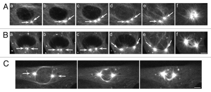
Figure 1.
Amphiastral spindle assembly in control BSC-1 cells. (A) The separation of the centrosomes is limited prior to NEB. The duplicated interphase centrosomes (part a, arrows) split apart but do not separate extensively (b–d, arrows). As the nuclear envelope breaks down (e and f), the bipolar spindle assembles. (B) The duplicated interphase centrosomes (part a, arrows) split and separate further apart (b–d, arrows). As the nuclear envelope breaks down (e and f), the bipolar spindle assembles. (C) The duplicated centrosomes (arrows) have migrated to opposite poles prior to nuclear envelope. GFP-α tubulin, fluorescence optics. Bar = 5 µm.

