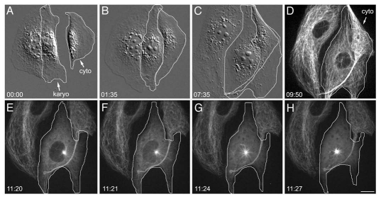
Figure 2.
Behavior of acentrosomal cell undergoing monopolar division. (A) MT organization following microsurgery. (A–C) Microsurgery and flattening of the karyoplast-cytoplast pair (surrounded by white outline). (D) Confocal fluorescence image of interphase karyoplast (white outline). There are MTs throughout the karyoplast. (E–H) Monopolar spindle formation. The single MT focus (E) is directly adjacent to the nuclear envelope. As the interphase MT network disassembles, cytoplasmic MTs are being drawn into the monopole. The nuclear envelope breaks down (F) and a monopolar spindle forms (H). GFP-α tubulin, fluorescence optics. Bar = 10 µm.

