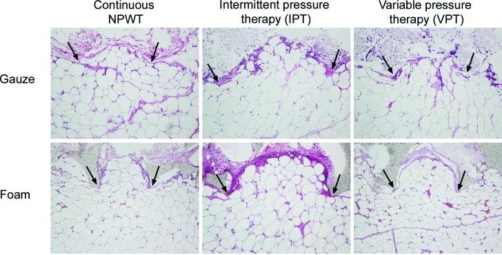
Figure 3.
Hematoxylin-eosin stained histological sections of wound biopsy specimens after 72 hours' continuous negative pressure wound therapy (NPWT), intermittent pressure therapy (IPT), or variable pressure therapy (VPT) using gauze or foam. The images show the wound filler (at the top of the images), the interface between the wound filler and the tissue, which was mainly composed of adipocytes (at the bottom of the images). It can be seen that both foam and gauze cause a repeating pattern of wound surface undulations, and the drawing of small tissue blebs into the pores of the foam and the spaces between the threads in the gauze, regardless of the mode of application of negative pressure. The protrusions of wound filler into the tissue are indicated by arrows.

