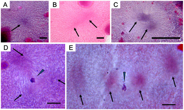
Figure 11.

Histological sections showing abnormalities in cold-shocked eggs after fertilization (af). A (80 min af) anuclear cell with one aster (arrow); B (90 min af) anuclear cell with two asters (arrows); C (100 min af) anuclear cell with two asters (arrows); D (120 min af) cell with a clumped nucleus (arrowhead) and a tripolar spindle with three asters (arrows); E (120 min af) cell with a clumped nucleus (arrowhead) and four asters (arrows). Scales denote 10 μm.
