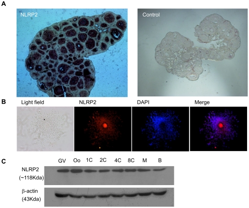
Figure 2. Developmental expression of NLRP2 protein in mouse.
(A) Immunohistochemical analysis of sequential sections from a 3-week-old mouse ovary using an anti-NLRP2 antibody. The original magnification was ×40. (B) Immunofluorescent detection of NLRP2 in cumulus–oocyte complexes after permeabilization and incubation with an anti-NLRP2 antibody. The original magnification was ×100. (C) Immunoblots of lysates isolated from oocytes and preimplantation embryos. Molecular masses (KDa) are indicated on the left; β-actin was used as a control.

