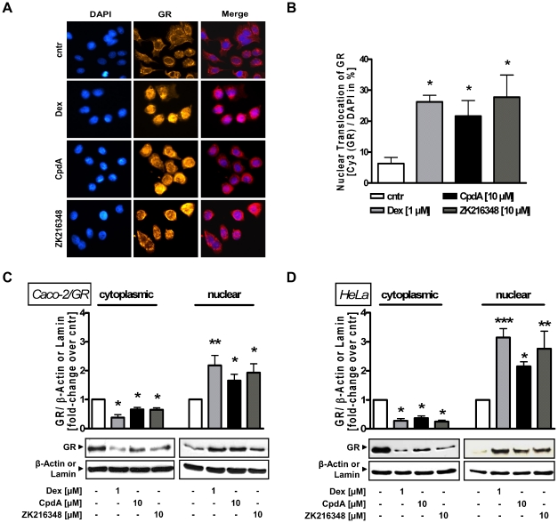
Figure 1. Effect of SEGRAs on GR binding and nuclear translocation.
(A) Immunofluorescence analysis for GR location in Caco-2 cells. Cells were treated for 3 h with Dex [1 µM], CpdA or ZK216348 [10 µM]. DAPI staining was used for visualisation of the cell nuclei. (B) 100 cells were randomly chosen, analysed and the percentage of nuclear GR calculated. Western blot analysis for translocation of GR in (C) Caco-2/GR and (D) HeLa cell lysates after 3 h cultivation with Dex or SEGRAs. One representative blot of three is shown. Densitometric analysis of GR is normalised to β-actin (cytoplasmic extract) or lamin (nuclear extract), respectively. Bars represent mean ± S.E.M., n = 3, *P<0.05; **P<0.01 relative to vehicle.

