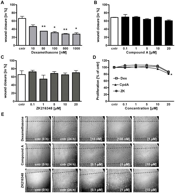
Figure 4. Effect of SEGRAs on intestinal epithelial cell restitution and proliferation.
IEC-6 cells were wounded and cultured in the presence of (A) Dex [0.1–1 µM] or (B) CpdA or (C) ZK216348 [1–20 µM] for 24 h. Cell migration was assessed using an in vitro migration assay. Bars indicate mean values of remaining wounded area ± S.E.M., n = 3, *P≤0.05, **P≤0.01 relative to control. (D) BrdU incorporation assay was used for determination of IEC-6 cell proliferation after 24 h incubation with Dex or SEGRAs [1–20 µM]. Results indicate mean ± S.E.M., n = 3, ***P≤0.001 relative to vehicle. (E) Representative experiments illustrating the effects of Dex and SEGRAs on intestinal epithelial cell restitution. Cntr (0 h) represents cells immediately after wounding, picture cntr (24 h) and others show wounds 24 h after cultivation with Dex, CpdA or ZK216348. Dotted line indicates original margin of wound (Magnification ×100).

