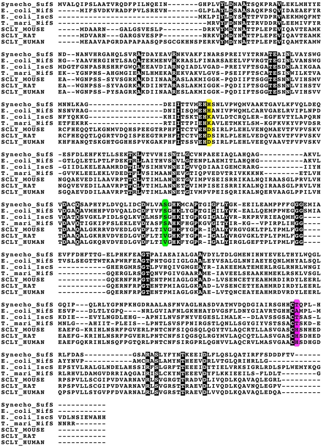
Figure 4. Alignment of representative sequences of bacterial SCL/CD enzymes (Synechocystis SufS, E. coli NifS, E. coli IscS and T. maritima NifS) and Mammalian Sec-specific SCLs (Mouse, Rat and Human).
Residue positions corresponding to D146, V256 and H389 (hSCL numbering) are indicated with a yellow, green and purple background respectively.

