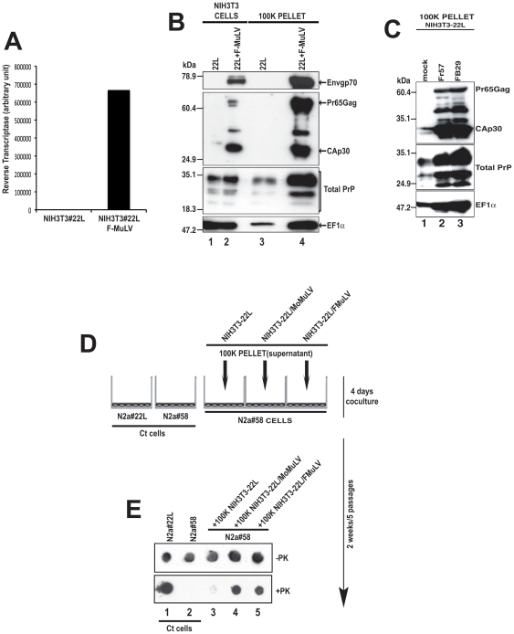
Figure 1. The Friend Murine Leukemia Virus (F-MuLV) strongly enhances PrP levels and prion infectivity released into the cell culture supernatant.
(A) Reverse transcriptase (RT) levels in NIH3T3-22L cells alone (22L) and NIH3T3 cells coinfected with the F-MuLV strain FB29 (22L+F-MuLV). (B) NIH3T3-22L (lane 1) and NIH3T3-22L/F-MuLV cells (lane 2) were assayed for F-MuLV expression using the DJ-39462 antibody for the viral envelope (Envgp70, Panel 1) and the DJ-39461 antibody for the viral capsid (CAp30, panel 2). PrP was detected using SAF-32 (Total PrP, panel 3) and exosomes using anti-EF1α (panel 4). The proteins recognized are indicated on the right side of the figure. Molecular mass markers are shown on the left side of the panel and lane numbers are indicated on the bottom of the figure. (C) NIH3T3-22L cells (mock) or NIH3T3-22L cells co-infected with the Friend viruses Fr57 or FB29 were analyzed by western blot for F-MuLV expression as well as PrP and exosome release as in panel B. Note that the F-MuLV Fr57 strain, like FB29, strongly enhances PrP and exosome release. Molecular mass markers are shown on the left side of the panel and lane numbers are indicated on the bottom of the figure. The proteins recognized are indicated on the right side of the figure. (D) Coculture assay. Uninfected target N2a#58 neuroblastoma cells grown on the bottom surface of a six well plate and a 0.4 mm pore size insert containing the 100 K pellet from the supernatants of NIH3T3-22L, NIH3T3-22L/MoMuLV or NIH3T3-22L/F-MuLV were cocultured for 4 days. The N2a#58 target cells were then passaged 5 times over 2 weeks and assayed for PrPSc. Ct = control. (E) Total PrP (-PK) and PrPSc (+PK) levels in N2a58 cells co-cultured with NIH3T3-22L/MoMuLV or NIH3T3-22L/F-MuLV supernatants were analyzed by dot blot. Ct cells = 22L infected and uninfected N2a#58 cells not exposed to supernatant. Lane numbers are shown at the bottom of the panel.

