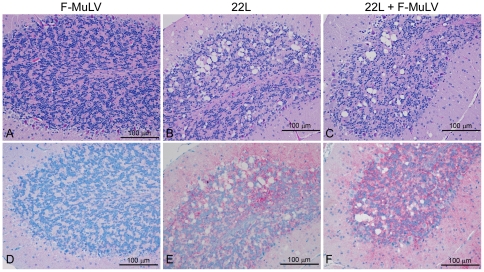
Figure 5. Vacuolation and PrPSc deposition in the brain is the same in mice infected with 22L or 22L plus FV.
(A–C) Hemotoxylin and eosin staining of the cerebellum from (B10 X A.BY)F1 mice inoculated (A) i.v. with FV alone, (B) i.p. with mouse 22L mouse scrapie alone, or (C) co-infected with 22L mouse scrapie i.p. and FV i.v.. Magnification = 200×. (D–F) PrPSc deposition in the cerebellum of (B10 X A.BY)F1 mice inoculated (D) i.v. with FV alone, (E) i.p. with mouse 22L mouse scrapie alone, or (F) co-infected with 22L mouse scrapie i.p. and FV i.v.. Sections were stained using the anti-PrP mouse monoclonal antibody D13 following hydrated autoclaving at high pressure and temperature. Red stain indicates the presence of PrPSc. Magnification = 200×.

