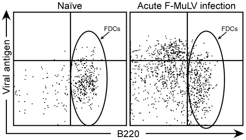
Figure 6. Analysis of follicular dendritic cells (FDCs) for Friend virus infection.
Flow cytometry was used to detect cell surface FV glycosylated gag using the monoclonal antibody mAb34 (Viral antigen). FDCs are B220+ cells gated for lack of expression of CD11c, CD8, CD19, CD4, CD3, CD11b and Gr-1, but staining positive for MHCII. The representative plot of FDCs from an acutely infected mouse shows no viral antigen staining above background (Naïve) levels.

