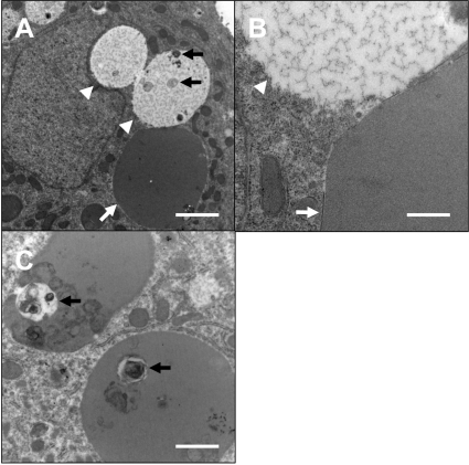
Fig. 3.
Ultrastructural findings. Gross appearance of a hepatocyte with inclusion and vacuoles (A) and higher magnification (B). Vacuoles were located adjacent to the nucleus and invaginated into it. The inclusions (white arrows) were surrounded by limiting membranes and composed of moderately electron dense, homogenous materials. On the other hand, the vacuoles (arrowheads) did not have obvious membranes and were filled with electron lucent, flocculent materials. Some vacuoles (A) and inclusions (C) contained debris of unknown origin (black arrows). Bars=2 µm (A), 0.5 µm (B) and 1 µm (C).

