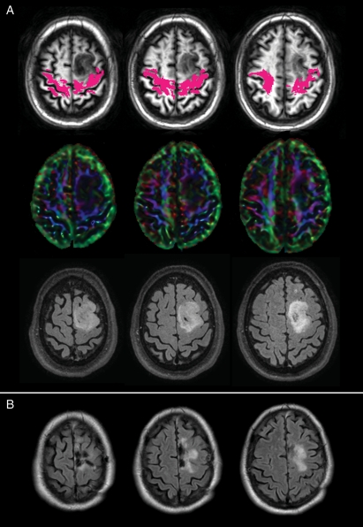
Fig. 1.
(A) A case of a left frontal oligodendroglioma infiltrating the left CST (magenta) both at the level of the subcortical white matter of the precentral gyrus and at the level of the centrum semiovale. Color maps show a reduction of anisotropy due to the presence of the lesion. Preoperative tumor volume was 29 cm3. (B) Postoperative MR shows a residual lesion in the area of deep infiltration of the fascicle. Involvement of CST is predictive of worse surgical outcome, as in patients with infiltrated CST it is less likely to obtain total resection (see Table 1).

