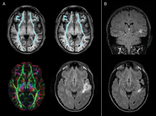
Fig. 2.
(A) A case of a left temporal oligodendroglioma with a deep nodule infiltrating the left IFO (cyan) at a posterior level where it passes from the occipital lobe to the external/extreme capsule. Color maps show a reduction of anisotropy due to the presence of the lesion; tractography shows a narrowing of the fascicle if compared to the contralateral normal IFO. Preoperative tumor volume was 27.5 cm3. (B) Postoperative MR shows the persistence of the deep nodule, resulting in a subtotal resection. Infiltration of IFO is predictive of worse surgical outcome, because in these patients total resection is less likely than is partial resection (see Table 1).

