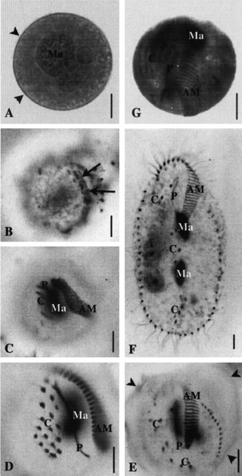
FIG. 2.
Excystment process (A to F) and effect of the E64 inhibitor (G) as observed on cells after silver proteinate impregnation and mounting in Citifluor (A and G) or dehydration and mounting in Canada balsam (B to F). Bars, 10 μm. (A to F) Excystment process. The resting cyst (A) is devoid of ciliature and enclosed in a wall (arrowheads). The macronucleus (Ma) is visible in the center of the cell. After induction of excystment, the first cortical event detected by silver proteinate impregnation is the appearance of a field of basal bodies (arrows in panel B). Thousands of basal bodies are quickly assembled and organized into anlagen (C and D), which will differentiate into specific ciliary structures of the young (E) and interphase (F) cell. Several of these anlagen are recognizable very early, and their maturation is easily monitored through the whole process. Adoral membranelles (AM) (or polykineties) and paroral membrane (P) (or haplokineties) represent the two sides of the oral ciliature in the interphase cell (F). The AM differentiate from the field of basal bodies already seen in panel B. This field enlarges (C); basal bodies are progressively organized into transversal rows (D) characteristic of the cell pattern (E and F). The paroral ciliature (P) of the vegetative cell (F) differentiates from a streak of basal bodies situated on the right of the AM anlage (C to E). The ventral cirri (C), which appear as dark points in the interphase cell (F), differentiate from streaks of basal bodies (C) which segment into individual cirri (D) and become pat-terned on the cortex as cell enlarges (E and F). The specific pattern of the interphase cell may be clearly recognized as the cell is still enclosed in its wall (arrowheads in panel E). During the process, the macronucleus elongates (D) and separates into two parts (E and F). (G) Effect of the E64 inhibitor. Inhibition of the excystment occurs as soon as ciliary anlagen are assembled, i.e., between stages C and D: anlagen of the oral apparatus (AM and P) as well as those of the cirri (C) are visible. The macronucleus (Ma) has not separated into two parts.

