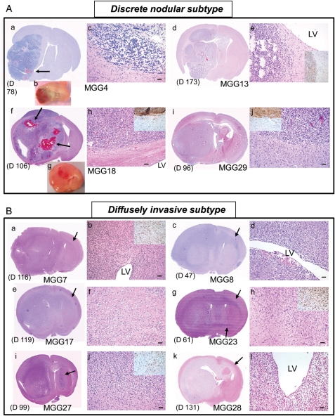
Fig. 3.
Invasiveness subtyping of GSC-derived GBM xenografts. (A) Discrete nodular subtype. a-c, MGG4. d, e, MGG13. f-h, MGG18. i, j, MGG29. H & E staining of coronal brain sections, except b and g, which show coronally cut planes of freshly removed brains. These tumors are characterized by relatively delineated borders between tumor mass and brain parenchyma, which was confirmed by immunohistochemistry for human-specific nestin (insets in e, h, and j). This tumor subtype was associated with increased vascularity (b, g) and intratumoral bleeding (arrows in a and f). There is typically no tendency for cells to accumulate in the subventricular zones (adjacent LV) (e, h). (B) Diffusely invasive subtype. a, b, MGG7. c, d, MGG8. e, f, MGG17. g, h, MGG23. i, j, MGG27. k, l, MGG28. These tumors are diffuse and highly invasive, and always extend to the contralateral hemisphere through white matter tracts (shown by arrows). Indistinct tumor-brain interface (f, h, and j; insets in b, h, and j showing human-specific nestin-positive cells on infiltrative edge) and heavy neoplastic infiltration in the subventricular zone (b, d, and l) are other phenotypic characteristics of this subtype of tumor. The timing of animal sacrifice when tumors caused significant symptoms is shown in days. (D) For each panel. LV, lateral ventricle. Bars, 50 µm.

