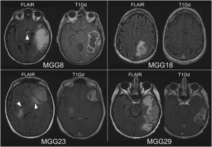
Fig. 4.
Patient preoperative MRIs. MR images taken before surgeries showing MGG8 and MGG23 that produced GSCs of highly invasive phenotype, and MGG18 and MGG29, from which GSCs of discrete nodular phenotype were derived. Arrowheads show hyperintense abnormal lesions in FLAIR images at the midline or bilateral regions suggestive of tumor infiltration. T1Gd, T1 images after infusion of gadolinium contrast agent.

