FIG. 5.
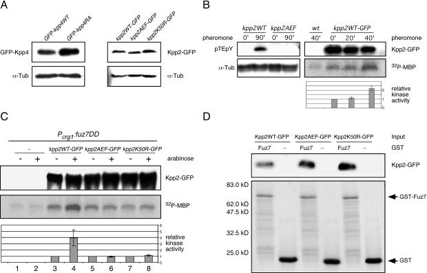
Pheromone as well as fuz7DD activates Kpp2 kinase activity. (A) Expression levels of kpp4 and kpp2 alleles constructed in this study. Left panel, Western analysis of protein extracts from SG200GFP-kpp4WT and SG200GFP-kpp4RA grown in CM-Glc by using anti-GFP antibody. In the lower panel the Western blot was stained with anti-α-Tub antibody as a loading control. Right panel, protein extracts from FB1kpp2WT-GFP, FB1kpp2AEF-GFP, and FB1kpp2K50R-GFP grown in CM-Glc were analyzed with anti-GFP antibody. In the lower panel the Western blot was stained with anti-α-Tub antibody as loading control. (B) Left panel, before and after pheromone stimulation, phosphorylation of Kpp2 was monitored with anti-pTEpY antibody. Extracts were prepared from FB1kpp2WT and FB1kpp2AEF 90 min after stimulation with synthetic a2 pheromone (90′) or after 90 min of DMSO treatment (0′). The upper panel shows a Western blot with anti-pTEpY antibody, and the lower panel shows a Western blot with anti-α-Tub antibody as a loading control. Right panel, Kpp2 kinase activity in FB2 (wild type [wt]) and FB2kpp2WT-GFP was assayed by MBP phosphorylation at 0, 20, and 40 min after pheromone addition. The upper panel shows precipitated Kpp2WT-GFP detected with anti-GFP antibody. The middle panel shows MBP phosphorylation, and the lower panel summarizes relative kinase activity measured by quantification of incorporated phosphate in three independent experiments. Error bars indicate standard deviations. (C) FB1 and the FB1-derived strains indicated on the top were shifted to arabinose-containing medium. Extracts were prepared prior to (−) and 90 min after (+) the shift. The upper panel shows precipitated Kpp2-GFP derivatives detected with anti-GFP antibody. The middle panel illustrates MBP phosphorylation, and the lower panel depicts relative kinase activity measured by quantification of incorporated phosphate in three independent experiments. (D) To demonstrate Kpp2 interaction with Fuz7 in vitro, protein extracts were prepared from strains expressing either kpp2-GFP, kpp2AEF-GFP, or kpp2K50R. These extracts were incubated with either GST-Fuz7 (Fuz7) or GST (−) bound to glutathione-Sepharose. The upper panel shows precipitated Kpp2-GFP (67 kDa) detected with anti-GFP antibody, and the lower panel illustrates GST fusion proteins (GST-Fuz7 [76 kDa] and GST [27 kDa]) bound to glutathione-Sepharose as detected by Coomassie blue staining. Experiments were performed twice with similar results.

