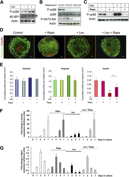
FIG. 5.
Leu represses endocrine progenitor cell development via mTORC1. A: Immunoblot for phospho (P)-rpS6 and 4E-BP1 expression in protein lysates from E13.5 pancreatic buds that were treated with Leu for 24 h. B: Immunoblot for P-rpS6, rpS6, and P-S473-Akt expression in protein lysates from E13.5 pancreatic buds that were treated with different concentrations of rapamycin (Rapa) for 24 h. C: Immunoblot for P-rpS6 expression in protein lysates from E13.5 pancreatic buds that were cultured with Leu in the presence or absence of rapamycin (10 mol/L) for 24 h. D: Immunohistological analyses of E13.5 fetal pancreata that were cultured for 7 days with and without Leu or rapamycin. Acinar cell and β-cell development were evaluated using an antibody against amylase (green) and an antibody against insulin (red), respectively. Nuclei were stained with Hoechst 33342 fluorescent stain (blue). Scale bar = 50 μm. E: Absolute areas that were occupied by nuclei and amylase- and insulin-positive cells were quantified using NIH Image J software. F and G: Real-time PCR quantification of insulin and Ngn3 mRNA in pancreata after 0, 1, 3, 5, and 7 days in culture without Leu (white bars), with rapamycin (gray bars), with Leu (black bars), and with both rapamycin and Leu (dashed bars). Data are mean ± SEM of at least three independent experiments. NS, not significant; *P < 0.05, **P < 0.01. (A high-quality digital representation of this figure is available in the online issue.)

