FIG. 2.
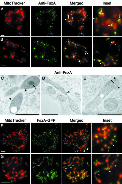
FszA formed belts and puncta within the mitochondria of Dictyostelium. (A and B) Immunofluorescence of Dictyostelium amoebae labeled with anti-FszA (green) and MitoTracker Red (red). (A) FszA formed belt-like (arrows) or punctate (arrowheads) structures in spherical mitochondria. (B) In rod-shaped mitochondria, one or two FszA foci were detected (arrowheads), often located near the ends of the organelles. Scale bar = 5 μm. (C to E) Immunoelectron microscopy with anti-FszA indicated that FszA (arrowheads) was located within the mitochondrion but did not concentrate within the electron-dense SMB (arrow) (C). Scale bars = 1 μm. (F and G) Expression of FszA-GFP in Dictyostelium. (F) At low expression levels, FszA-GFP was targeted to the mitochondria where it mostly formed a single belt in each spherical organelle (arrowhead). (G) At high expression levels, FszA-GFP formed extended helices (arrowheads) in long, tubular mitochondria. Scale bar = 5 μm.

