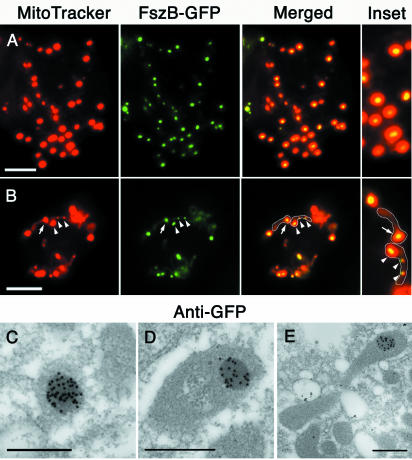
FIG. 3.
FszB-GFP localizes to an SMB in Dictyostelium amoebae. (A and B) FszB-GFP accumulated within mitochondria to regions called SMBs that stained brightly with MitoTracker Red. SMBs are present in spherical (A) and tubular (B) organelles. (B) Mitochondria have been outlined. Usually, a single SMB was detected near the end of tubular mitochondria (arrow), but occasionally one or two additional SMBs are also visible within an organelle (arrowheads). (A and B) Scale bars = 5 μm. (C to E) Immunoelectron microscopy with anti-GFP gold demonstrates that FszB-GFP almost exclusively localized to the electron-dense SMB and in mitochondrial tubules this structure was often near one end (E). Scale bar = 0.5 μm.

