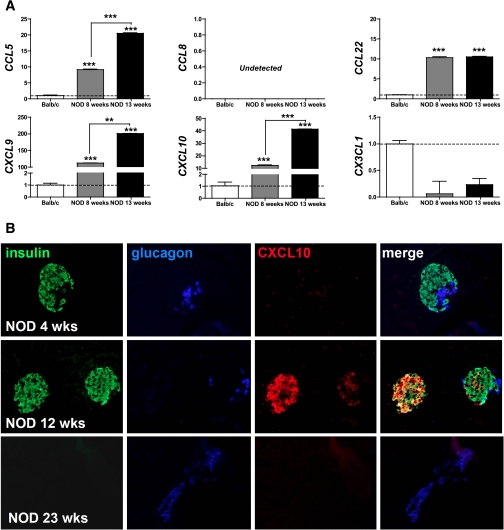
FIG. 5.
Chemokine expression in the NOD model of spontaneous type 1 diabetes. A: Chemokine mRNA transcript expression was quantified in islets isolated from Balb/c (n = 6) as well as 8- and 13-week-old female NOD mice (n = 6) by qRT-PCR. Data were quantified using the 2–Δ Δ CT method expressed as means ± SD (n = 3) and normalized to housekeeping Hprt and Balb/c samples (calibrator). Relative chemokine mRNA expression is displayed in relation to Balb/c samples (dotted line); asterisks indicate significant differences between control and cytokine-treated islets. B: CXCL10 production by pancreatic β-cells as a function of female NOD age was determined as detailed in RESEARCH DESIGN AND METHODS. Note the weak CXCL10 staining in acinar tissues in 4-week-old NOD mice, preferential colocalization with insulin in 12-week-old NOD mice, and complete absence of CXCL10 at 23 weeks of age. (A high-quality digital representation of this figure is available in the online issue.)

