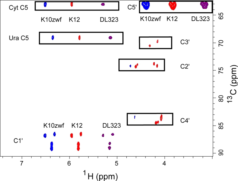
Fig. 2.
2D non-constant time HSQC spectra of all four labeled nucleotides extracted from K10zwf (blue contours, left shifted) or K12 (red contours) or DL323 (purple contours, right shifted) E. coli strains grown on [3-13C]-pyruvate. Growth on [3-13C]-pyruvate results in label at only C1′ and C5′ for DL323 and K10 strains, whereas growth of K12 leads to some residual label at C2′, C3′, and C4′ in addition to the major labels at the C1′ and C5′ carbons. The cytosine (Cyt) and Uracil (Ura) C5 resonances at 96.67 and 102.69 ppm respectively are folded into the spectrum

