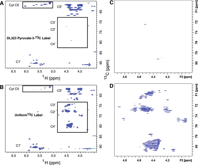
Fig. 4.
NMR spectra showing enhanced resolution afforded by site selective labeling of A-Site RNA bound to paromomycin. The experiments were performed on the A-Site RNA a site-selectively 13C-GTP and CTP labeled and b uniformly 13C-GTP and CTP labeled. 2D non-constant time HSQC spectra of the ribose region. The cytosine (Cyt) and Uracil (Ura) C5 resonances at 96.67 and 102.69 ppm respectively are folded into the spectrum and are boxed to highlight the reduced signal from the uniformly labeled sample. The C2′, C3′, and C4′ regions are boxed to highlight the absence of labeling in selectively labeled sample. Blown-up views of the C2′, C3′, and C4′ regions boxed in (a) and (b) are depicted in (c) and (d) respectively

