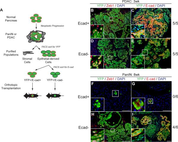
Figure 5. Epithelial and mesenchymal states are plastic.
(A) Schematic of orthotopic transplantation experiments. (B–E) Fluorescent images taken 3 wks following transplantation of YFP+ cells from PDAC mice into NOD/SCID hosts. Tumors form in all mice regardless of E-cad status (n=5 for each condition). YFP+E-cad+ and YFP+E-cad− cells are present in both conditions (C and E), as are YFP+Zeb1+ and YFP+Zeb1− cells (B and D). (F–I) Fluorescent images taken 8 wks following transplantation of YFP+ cells from PanIN mice into NOD/SCID hosts. After transplantation of YFP+E-cad+ cells, no tumors are found (n=6); the few transplanted YFP+ cells that remain are Zeb1− and E-cad+ (F, G). Transplantation of YFP+E-cad− cells results in tumor formation (H, I). Tumors contain both E-cad+ and E-cad− cells (I) as well as Zeb1+ and Zeb1− cells (H), providing direct evidence for MET.

