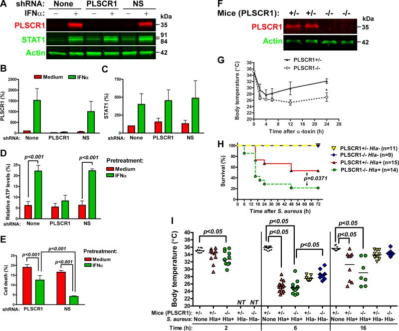
Figure 4.
Expression of PLSCR1 is necessary for INFα-induced protection from α-toxin. A549 cells were stably transfected with PLSCR1-specific or non-silencing (NS) shRNA vectors and analyzed for expression of PLSCR1 and STAT1 by immunoblotting. A representative immunoblot (A) and densitometry analyses of PLSCR1 (B) and STAT1 (C) expression normalized to actin are shown (mean±SD of three independent experiments). A549 cells stably expressing PLSCR1-specific or NS shRNA were pretreated with IFNα and treated with 2.5 μg/ml α-toxin. (D): Relative ATP levels remaining after 16-h treatment with α-toxin. The data are mean±SD of three independent experiments. (E): Cell death after 24-h treatment with α-toxin. The data are mean±SD of quadruplicate cultures and are representative of three experiments. (F): Immunoblot analysis of PLSCR1 expression in the lungs of littermate PLSCR1+/- and PLSCR1-/- mice. (G): Littermate PLSCR1-/- and PLSCR1+/- mice were administered α-toxin via intranasal route. Body temperature was measured at the indicated time points. Asterisk indicates statistically significant difference in body temperature between the groups at 24 h after α-toxin (mean±SEM, p<0.05, n=4). (H): Littermate PLSCR1-/- and PLSCR1+/- mice were infected with 2.5×10⁸ CFU/mouse of α-toxin-producing Hla+ S. aureus or 3.65×10⁸ CFU/mouse of isogenic α-toxin-deficient Hla- S. aureus and monitored for the signs of moribund condition for up to 72 h. (I): Body temperature for control non-infected mice and S. aureus-infected mice at 2, 6 and 16 h post infection is shown (median and individual values, NT - not tested).

