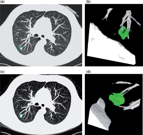
Figure 1.
Baseline and 3-month follow-up CT images in a 68-year-old participant of the NELSON study. Transverse thin-section CT (a,c) and volume-rendered reconstruction (b,d) images show a lobulated pulmonary nodule with vessel attachment (boxed on a,c and green area in b,d). On the baseline scan (a,b) the volume was 303 mm3. On the 3-month follow-up CT (c,d), the volume was 576 mm3. This is consistent with a percentage volume growth of 90% and a volume-doubling time of 98 days. Histopathology of the resected nodule: squamous cell carcinoma.

