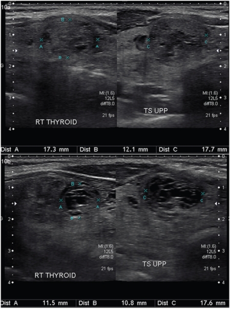
Figure 10.
Temporal evolution of a benign thyroid nodule. (a) A solid nodule with few cystic spaces and hypoechoic halo is demonstrated in the longitudinal and transverse planes. (b) The follow-up US images of the same nodule obtained after 1 year shows an increase in the cystic degeneration of the nodule with a slight decrease in its size.

