Fig 5.
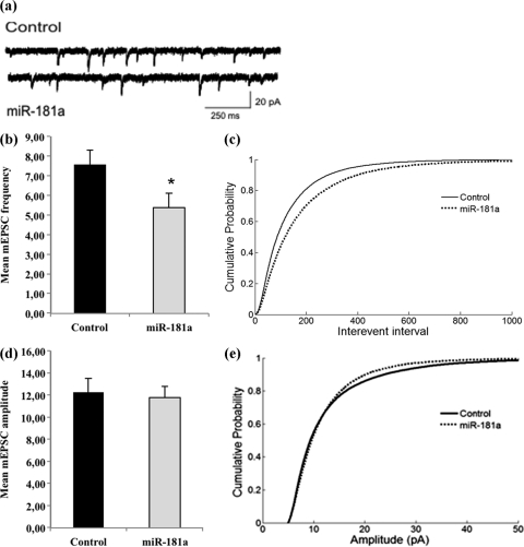
miR-181a reduces the frequency of mEPSCs in hippocampal neurons. Neurons were transfected as described for Fig. 4 and subjected to patch-clamp recordings between 17 and 19DIV. (a) Representative current traces from neurons transfected with either miR-181a or a control RNA. (b) Average mEPSC frequencies of neurons transfected either with miR-181a or a control RNA. n = 13 neurons per condition, originating from three independent transfections. P < 0.05. (c) Cumulative probability of interevent intervals from neurons analyzed in panel b. n = 26224 (control), 18285 (miR-181a). P < 0.001 by Kolmogorov-Smirnov (KS) test. (d) Average mEPSC amplitudes of events from neurons analyzed in panel b. P = 0.78. (e) Cumulative probability plot of amplitudes of events from neurons analyzed in panel b. n = 26309 (control), 18388 (miR-181a). P < 0.001 by KS test.

