
Figure 3.
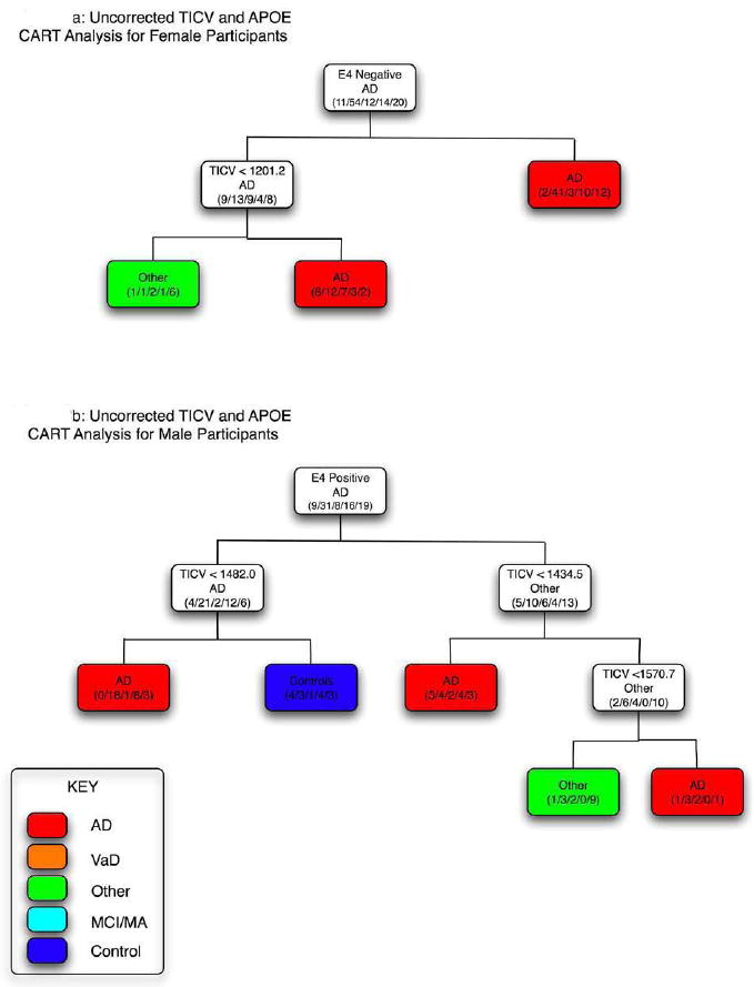
Classification and Regression Tree (CART) analysis by total intracranial volume (TICV) and presence of the ε4 allele for females (a) and males (b). As in Figure 2, when the criterion heading for each line is met, the direction is to the left. Accordingly, there is a cluster (asterisk) where Alzheimer disease (AD) subjects had brain volume ≤ 1482.04 and no controls were in this branch. No branching occurred by TICV that appeared meaningful in females, as the CART analysis classified AD strictly by apolipoprotein E (APOE) genotype on the first branch and those who were ε4+ (right branch) were not further subdivided by TICV. Likewise almost all other subjects who were classified by the ε4- condition had TICV > 1201.2 cm3.