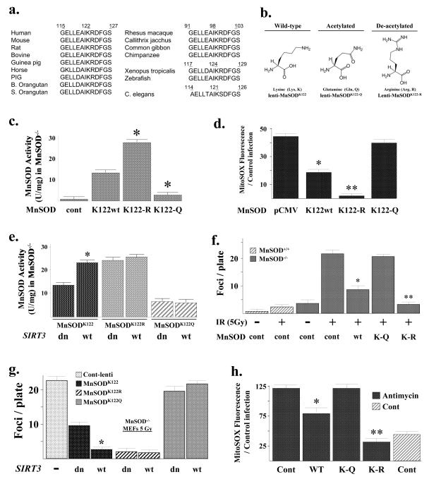
Figure 5. MnSOD contains an evolutionarily conserved lysine residue that regulates SOD activity.
(a) Multiple species contain a potentially reversibly acetylated lysine residue. The MnSOD protein sequence from multiple species was BLASTed based on the reversibly acetylated lysine located at amino acid 122 in mice. A 13-amino acid motif (GELLEAIK*RDFGS) was identified that is present in multiple species. (b) Substitution of a lysine with a glutamine (Q) mimics an acetylated amino acid state, while substitution with an arginine (R) mimics a deacetylated amino acid state (Li et al., 2007; Schwer et al., 2006). (c) MnSOD lysine acetylation status directs dismutase activity. MnSOD−/− MEFs were infected with a control lentivirus or lenti-MnSODwt, lenti-MnSODK122-Q, or lenti-MnSODK122-R. Twenty-four hours after infection, MnSOD activity was determined as outlined above (Fig. 3 legend). (d) Mitochondrial superoxide levels are decreased in cells overexpressing a MnSODK122-R mutant gene. MnSOD−/− MEFs were infected with the various MnSOD lentiviruses and superoxide levels were determined as described above. (e) MnSOD−/− MEFs were infected with the wild-type and mutant MnSOD lentiviruses with either lenti-Sirt3-wt or lenti-Sirt3-dn and assayed for MnSOD activity. (f) The MnSODK122-R mutant decreases IR-induced contact inhibition in the MnSOD knockout MEFs. MnSOD−/− MEFs were infected with the MnSOD lentiviruses, plated at 1 × 10⁶/100 mm dish, exposed to 5 Gy IR, followed by long-term culture (28 days), staining with crystal violet, and measurement of foci formation. (g) MnSOD−/− MEFs were infected with the wild-type and mutant MnSOD lentiviruses with either lenti-Sirt3-wt or lenti-Sirt3-dn and measured for foci formation as above. (h) Mitochondrial superoxide levels in MnSOD−/− MEFs exposed to Antimcyin A are decreased by infection with lenti-MnSODK122-R, Sirt3−/− MEFs were treated with 5 μM of Antimycin A for 3 hours and mitochondrial superoxide levels were determined as described above. Results for all the panels in this figure are the mean of at least three separate experiments and error bars represent one standard deviation. * indicates P < 0.05 and ** indicates P < 0.01 by t-test.

