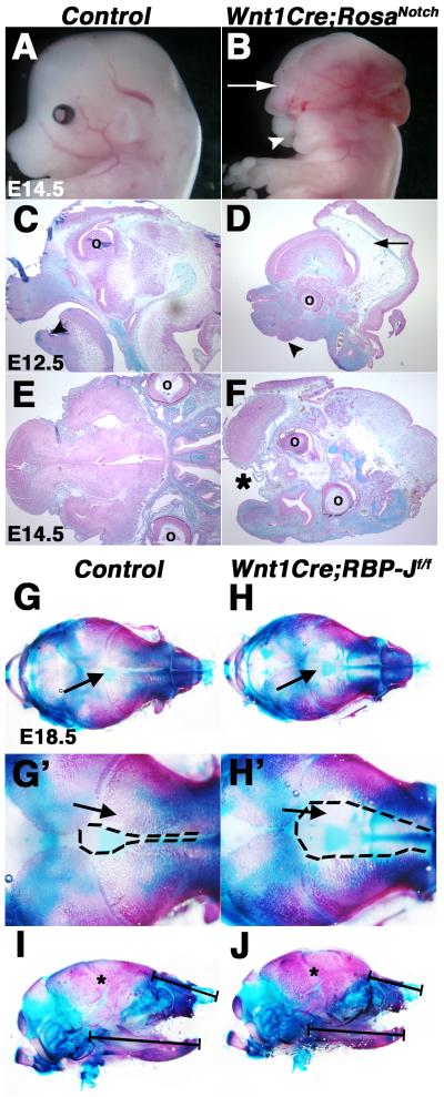
Fig. 1.
Craniofacial malformations result from activation or inactivation of Notch signaling in the neural crest. (A-F) Wnt1Cre;RosaNotch embryos (n=4) exhibit craniofacial malformations, consisting of exencephaly (B, D, arrows), micrognathia (D, arrowhead), cleft face (B, arrowhead) and palate (F, asterisk). Embryos are shown in whole mount (A,B) and in Alcian blue and nuclear fast red counter stained cranial sections (C-F). (G-H’) Alcian blue and Alizarin red stained cranial skeletal preparations of E18.5 Wnt1Cre;RBP-Jf/f embryos exhibit expanded frontal cranial sutures as a result of deficient formation of craniofacial frontal bones apparent in reduced Alizarin red-stained structures, as compared to a wildtype control littermate (arrows) (n=4). (I-J) Mandible size and nasal cartilage are reduced in Wnt1Cre;RBP-Jf/f embryos (n=6), as compared to a wildtype control littermate (n=6). ° denotes eye placement. Dotted lines (G’, H’) represent the area of proximal frontal bones. * indicate apparently normal coronal suture closure (I,J). Black brackets (I, J) denote length of mandible and nasal cartilage.

