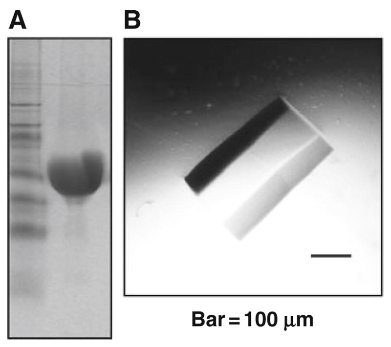
Fig. 24.1.

Assessment of the homogeneity of NBD1 preparations. (a) Coomassie blue stained SDS-Page polyacrylamide gel of human WT-NBD1 (aa 389–673), left lane marker, right lane 50 μg of human WT-NBD1, indicating it is the predominant protein in the preparation (see Section 3.1). (b) Crystal of murine F508del-NBD1, with no further mutations (aa 389–673), again, suggestive of high homogeneity of preparation (see Section 3.2).
