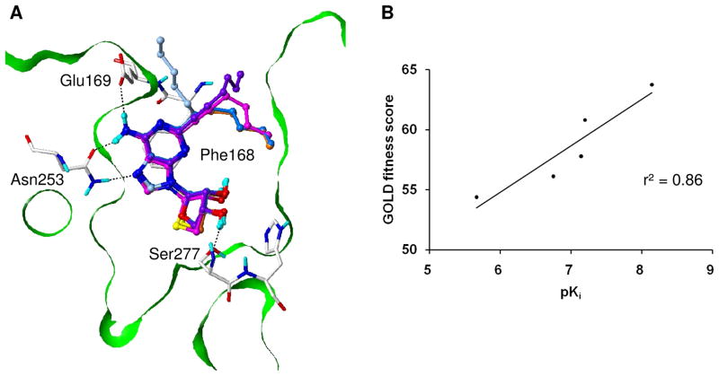
Figure 6.
Predicted binding modes of the five C2-substituted and N6-unsubstituted derivatives in an hA2AAR crystal structure,23 and the correlation between their experimental binding affinities and docking scores. (A) The adenine and sugar moieties of the molecules showed almost exactly the same binding modes maintaining the key interactions. Compounds 4a, 4f, 4g, 4l, and 4m are depicted as ball-and-stick with carbon atoms in purple, light-blue, magenta, skyblue and orange, respectively. (B) The scatter plot of the pKi values and GOLD fitness scores for the five compounds showed a good correlation with r2 of 0.86.

