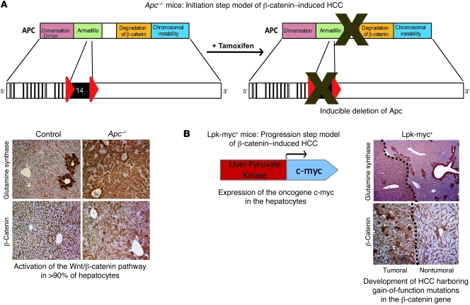
Figure 1. Description of the different genetically modified mouse models harboring oncogenic β-catenin activation in the liver.
(A) Initiation step model of β-catenin–induced HCC. In this previously described model (14, 15), Apc inactivation in nearly all hepatocytes of the liver lobule is induced by tamoxifen injection and is evidenced by glutamine synthetase, cytosolic, and nuclear β-catenin stainings. Apcflox/flox, TTR-Cre-Tam mice are referred to as Apc–/– mice and Apcflox/flox, Cre-negative mice are control mice. (B) Progression step model of β-catenin–induced HCC. In this previously described model (9, 16), expression of the c-myc oncogene is targeted in the hepatocytes under the control of the Lpk gene and leads to the development of HCCs that harbor activated β-catenin mutations (9, 16) and stain positive for glutamine synthetase and nuclear β-catenin.

