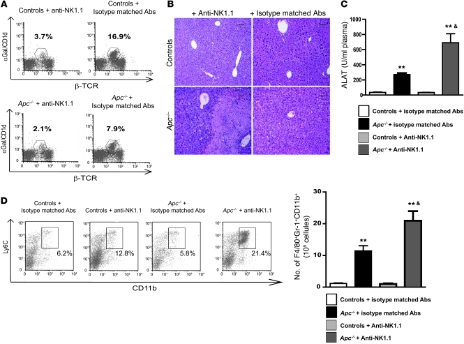
Figure 7. iNKTs display antiinflammatory properties during β-catenin–induced liver inflammation.
(A) FACS analysis of NPCs was performed to assess iNKT cell depletion in controls and Apc–/– livers after treatment with anti-NK1.1 or isotype-matched irrelevant antibodies 8 days after tamoxifen injection. (B) Paraffin-embedded sections obtained from controls and Apc–/– livers after treatment with anti-NK1.1 or isotype-matched irrelevant antibodies were stained with H&E. Scale bars: 50 μm. (C) Serum transaminase levels (ALAT) were measured in controls and Apc–/– mice after treatment with anti-NK1.1 or isotype-matched antibodies. (D) The sub-populations of F4/80+CD11b+Ly6C+ immature macrophages were quantified in the livers of Apc–/– and control mice treated with either anti-NK1.1 or isotype-matched irrelevant 8 days after tamoxifen injection. Dot plots show representative FACS analyses of immature macrophages, gated on F4/80+ cells. The numbers indicate the percentage of these cells among the F4/80+ cells. All data are representative of 3 independent experiments with 4 mice/group. **P < 0.01 (controls vs. Apc–/–); &P < 0.01 (Apc–/– + isotype-matched Abs versus Apc–/– + anti-NK1.1 Abs).

