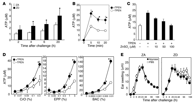
Figure 2. Zn deficiency increases ATP release by keratinocytes in response to irritant chemicals.
(A) 1% CrO was painted on the ears of ZA (white bars) and ZD (black bars) mice (n = 5), and skin samples were taken at the indicated time points. ATP secretion from skin organ cultures was quantified. (B–D) Pam-212 keratinocytes were treated with (B and C) 0.3% CrO or (D) titrated concentrations of CrO, EPP, or BAC in the presence (black circles/bars) or absence (white circles/bars) of 2 μM TPEN. ATP in the culture supernatants was quantified at (B) the indicated time points or (C and D) 10 minutes after stimulation. (C) Cells were cultured with 10–100 μM ZnSO4. *P < 0.05, compared with cells that were not treated with TPEN. (E) ICD responses to CrO were induced, as described in Figure 1. ZA and ZD mice (n = 5) received local injections of potato apyrase on the right ear (black circles) or PBS alone on the left ear (white circles) before and after CrO application to both ears. The data shown are the swelling responses (mean ± SD). *P < 0.05, between the apyrase and PBS treatments. Data are representative of 3 independent experiments.

