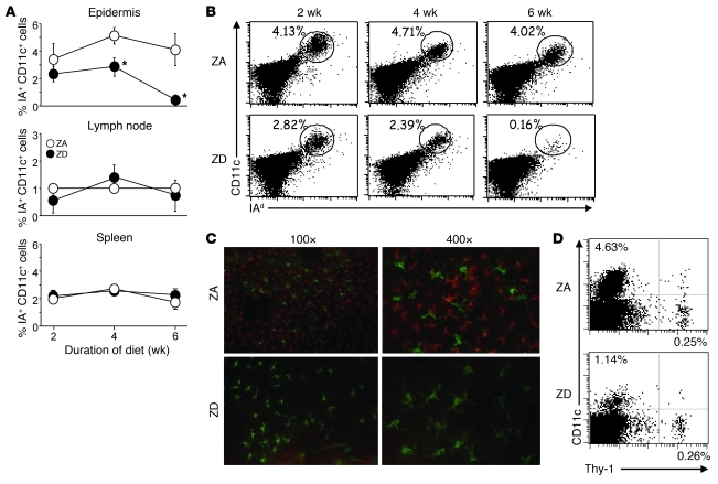
Figure 4. Loss of epidermal LCs in Zn deficiency.
(A) Cell suspensions of epidermis, axillary, and inguinal lymph nodes and spleens were prepared from mice fed ZA (white circles) or ZD (black circles) diets for the indicated time and stained for I-A and CD11c antigens. The percentages of I-A and CD11c double-positive cells were assessed within each live-gated cell population. Results are the mean ± SD (n = 3). *P < 0.05, compared with mice fed the ZA diet. A representative FACS analysis of live-gated epidermal cell suspensions from mice fed ZA or ZD diets for 6 weeks and stained for (B) I-A and CD11c or (D) Thy-1 and CD11c antigens. Numbers indicate the percentages of cells in the (B) circles or (D) gate. (C) Immunofluorescence of epidermal whole mounts stained for I-A (red) and Thy-1 (green) from mice fed ZA or ZD diets for 6 weeks. Original magnification, ×100 (left panels); ×400 (right panels). Data are representative of 3 independent experiments.

