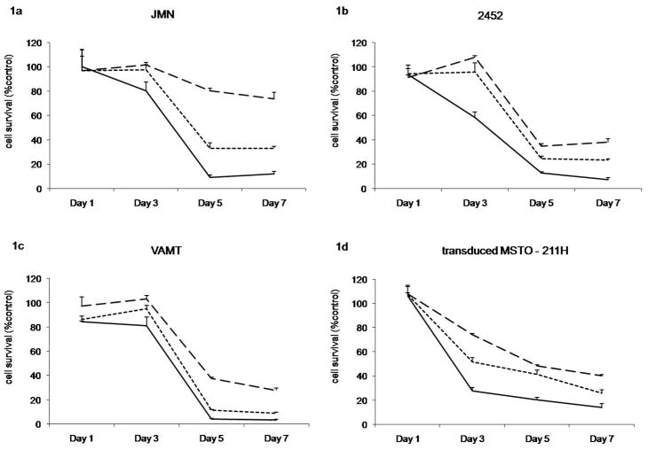
Figure 1. Cytotoxicity of NDV(F3aa)GFP against mesothelioma cell lines in vitro.
LDH assays of mesothelioma cell lines of (a) JMN, (b) H-2452, (c) VAMT, and transduced (d) MSTO-211H* at different MOIs (0.01=dashed line, 0.1=dotted line, 1=solid line) at day 1, 3, 5, 7. Data are expressed as the ratio of surviving cells determined by comparing the LDH-concentration of infected sample relative to control untreated cells.

