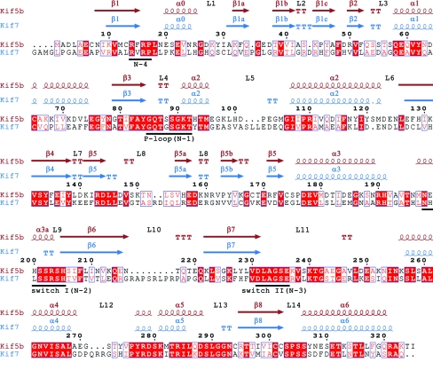
Figure 4.
Sequence alignment and secondary-structure elements derived from the crystal structures of the Kif5b (conventional kinesin, kinesin-1; PDB entry 1bg2; Kull et al., 1996 ▶) and Kif7 motor domains. The numbering relates to residues of Kif5b. Identical residues are coloured in white on a red background; similar residues are shaded in red. The ATP-binding pocket and the switch regions (N1–N4) are underlined in black.

