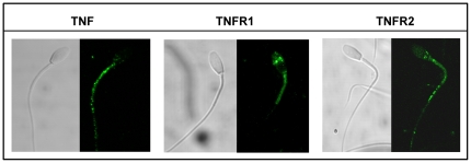
Figure 2. Subcellular distribution of TNF alpha and TNR receptors R1 and R2 in stallion spermatozoa.
Their subcellular distribution in fixed and permeabilized stallion spermatozoa was assessed by immunocytochemistry with specific antibodies as described in material and methods. TNF was localized in the mid piece and rest of the tail, TNFR1, was present in the acrosomal region and mid piece, while TNFR2 was present in the post acrosomal region, mid piece and rest of the tail. All images were obtained with a Bio Rad MRC1024 confocal microscope. Magnification, 60x.

