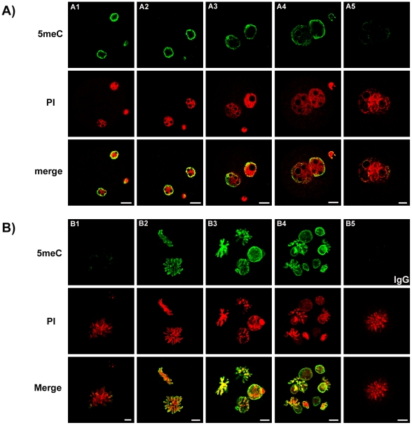
Figure 1. Pattern of anti-5meC staining in early preimplantation stage embryos.
Zygotes were collected directly from the oviduct 16 h – 25 h after the ovulatory injection of hCG. Images show zygotes at PN stage 1-5 (A1-5) and condensing chromosomes (B1), 2-cell (B2), 4-cell (B3) and 8-cell (B4). Mitotic zygotes stained with non-immune IgG control is also shown (B5). Antigenic unmasking of 5meC (green) was by brief acid exposure. DNA was counter stained with propidium iodide (PI, Red). Images from these two channels were merged to show co-localization (merge). Images of zygotes were single-equatorial confocal sections (0.77 µm), images of nuclei from 2-cell to 8-cell stage were Z-stacks of multiple sections through each nuclei of the embryo. The images shown here are representative of at least seven independent replicate experiments with at least 10 embryos per observation group per replicated. The scale bars are 10 µm.

